Offer ends October 18.
This is some text inside of a div block.
Restart a Heart BLS Sale
RESTART99
Copy code

Microscopy involves studying tiny objects using an instrument that magnifies images of items too small to be seen with the naked eye. Beyond the standard types, there are several specialized forms of microscopy, such as:- **Electron Microscopy**: Utilizes an electron beam instead of light to create images.- **Fluorescence Microscopy**: Relies on the property of fluorescent materials to emit visible light when exposed to ultraviolet (UV) rays.- **Immune Electron Microscopy**: Involves examining biological specimens with bound specific antibodies using electron microscopy.- **Immunofluorescence Microscopy**: Uses antibodies tagged with a fluorescent substance, along with a fluorescence microscope, to reveal antibody binding through visible light emitted under UV exposure.- **Nomarski Microscopy**: Employs a specialized optical system (Nomarski optics) to perform "differential interference contrast microscopy."- **Time-lapse Microscopy**: Captures images of the same object at set intervals to observe processes like cell division over time.The term "microscopy" is derived from Greek words: "mikros," meaning small, and "skopeo," meaning to view, thus translating to "viewing small objects."
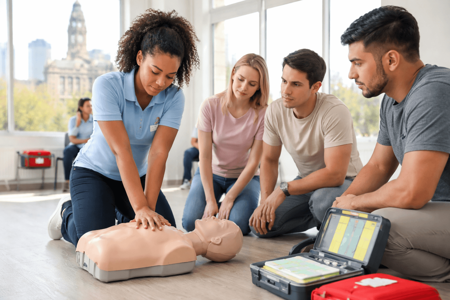
Our technology delivers immediate, actionable feedback that refines your skills in real time.
Understand how to safely and effectively operate an Automated External Defibrillator (AED) for adults, children, and infants.
Learn how to perform the Heimlich maneuver and clear airway obstructions caused by choking in both adults and infants.
Gain experience working as part of a team when more than one rescuer is available during an emergency.
Receive your AHA CPR Certification E-Card on the same day you complete the class and pass the exam.

CPR Certification Labs provides professional CPR and emergency training courses based on recognized industry standards.