Offer ends July 19.
This is some text inside of a div block.
It’s Hot. Skip the Classroom
SUMMER99
Copy code
Computerized axial tomography involves creating images of internal body structures using a computer that processes data from multiple images to form pictures. The CAT scan can detect soft tissues and other features that are not visible with standard X-rays. With the same radiation dose as a regular X-ray machine, it can produce images of body slices with about 100 times greater clarity. The tomograms for a CAT scan are typically spaced 5 or 10 mm apart. The CAT machine rotates 180 degrees around the patient, which is why it is called "axial." It emits a narrow X-ray beam from 160 different positions. Crystals positioned opposite the beam capture and record how the tissue and bone absorb the X-rays. This data is sent to a computer, which converts it into a two-dimensional cross-sectional image. The invention of CAT scanning in 1972 is credited to British engineer Godfrey N. Hounsfield (later knighted) and South African (later American) physicist Alan Cormack. By 1979, CAT scanning was widely used, and Hounsfield and Cormack were awarded the Nobel Prize in Medicine for their work. The CAT scan is also referred to as the CT (computerized tomography) scan.
Our technology delivers immediate, actionable feedback that refines your skills in real time.
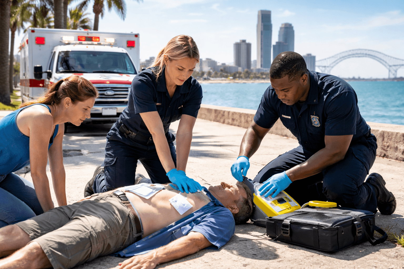
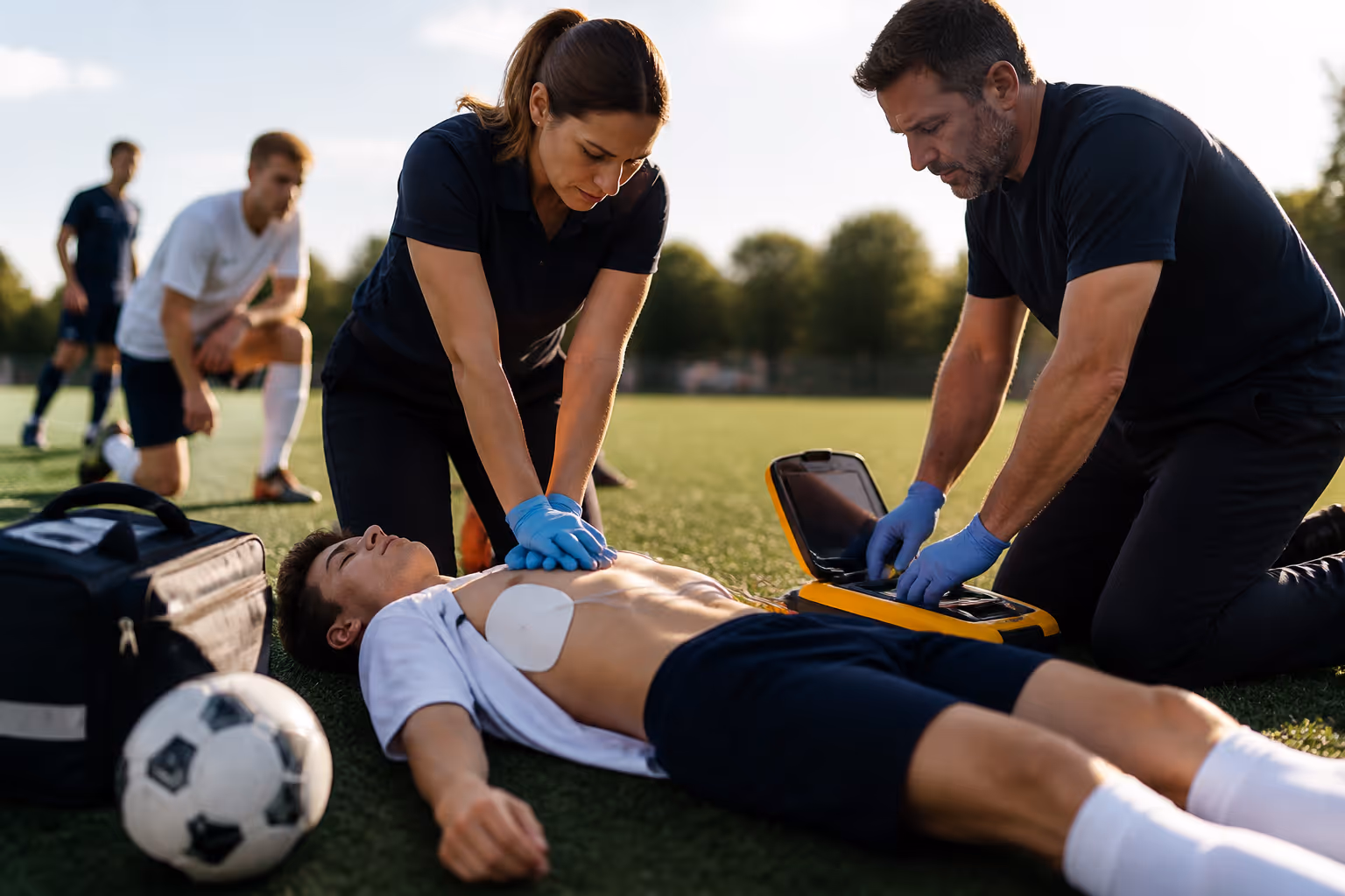
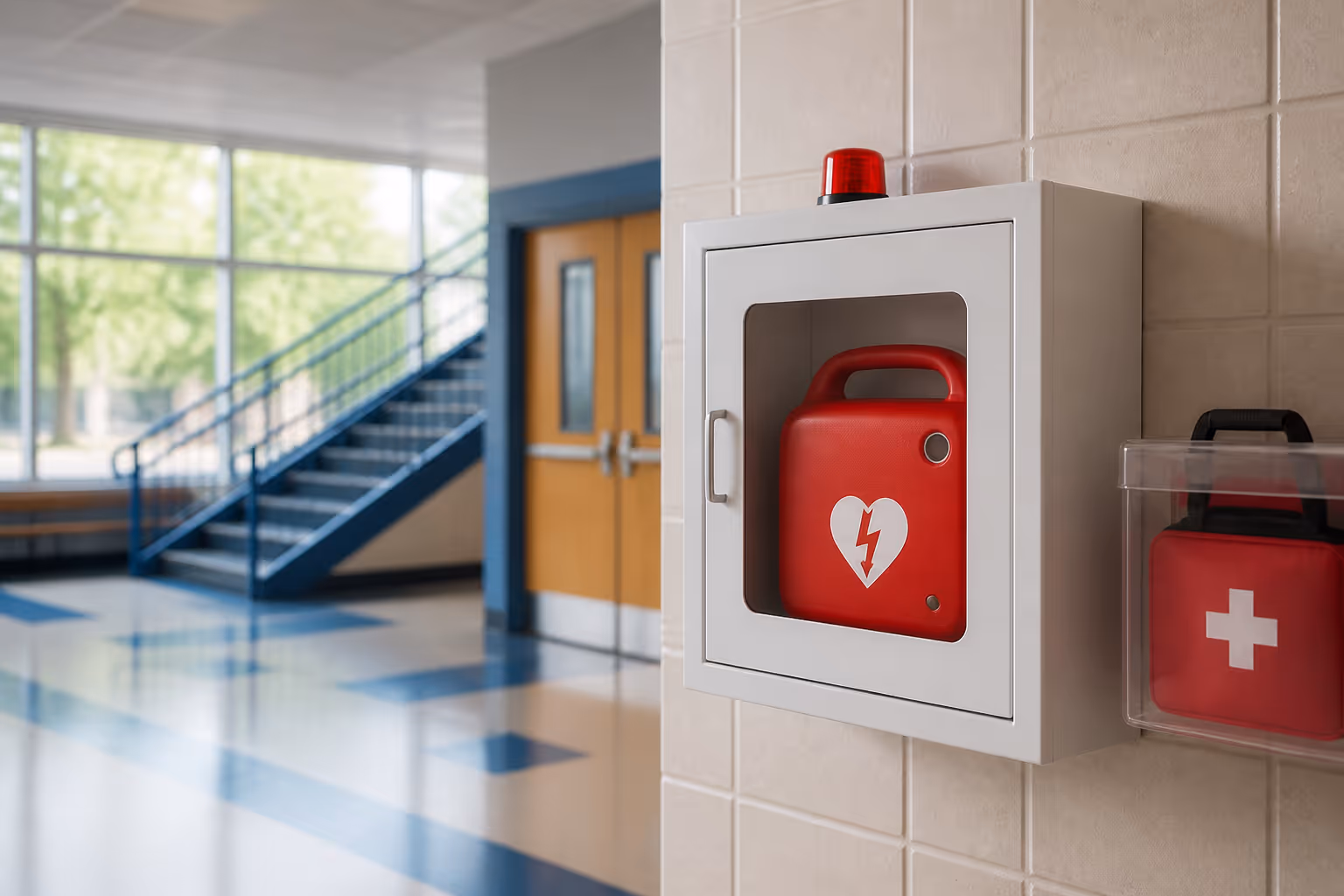
Understand how to safely and effectively operate an Automated External Defibrillator (AED) for adults, children, and infants.
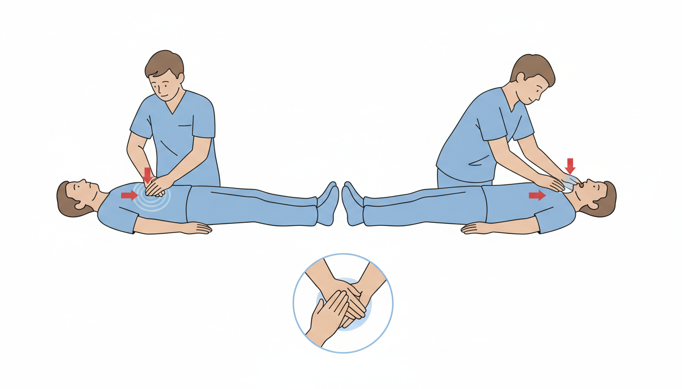
Learn how to perform the Heimlich maneuver and clear airway obstructions caused by choking in both adults and infants.
Gain experience working as part of a team when more than one rescuer is available during an emergency.
Receive your AHA CPR Certification E-Card on the same day you complete the class and pass the exam.


CPR Certification Labs provides professional CPR and emergency training courses based on recognized industry standards.